References
With a little lurid hilighting and graphics.
All images are taken from their respective papers.
Use of V1 in visualization:
1.
Transient Activity in the Human Calcarine Cortex During Visual-Mental
Imagery: an Event-Related fMRI Study.
Klein, Paradis, Poline,
Kosslyn, Le Bihaan, 1997.
Abstract: Although it is largely accepted that visual-mental imagery and perception draw on many of the same neural structures, the existence and nature of neural processing in the primary visual cortex (or area V1) during visual imagery remains controversial. We tested two general hypotheses: The first was that V1 is activated only when images with many details are formed and used, and the second was that V1 is activated whenever images are formed, even if they are not necessarily used to perform a task. We used event-related functional magnetic resonance imaging (ER-fMRI) to detect and characterize the activity in the calcarine sulcus (which contains the primary visual cortex) during single instances of mental imagery. The results revealed reproducible transient activity in this area whenever participants generated or evaluated a mental image. This transient activity was strongly enhanced when participants evaluated characteristics of objects, whether or not details actually needed to be extracted from the image to perform the task. These results show that visual imagery processing commonly involves the earliest stages of the visual system.
2. Visual Angle of the Mind's Eye Before and After Unilateral Occiptal Lobectomy.
Farah
MJ, Soso MJ, & Dasheiff RM
Abstract: Do mental images occur
in a spatially mapped (i.e., analog, or array-format)
representational medium? Kosslyn's (1978) method was used to measure
the visual angle of "the mind's eye" to estimate the extent
of the imagery medium before and after unilateral occipital
lobectomy. It
was found that the overall size of the largest possible image was
reduced following the surgery. In addition, only the horizontal
extent, and not the vertical extent, of the imagery medium was
reduced. Finally, it was determined
that the S[ubject] understood the tasks, was not aware of our
predictions, and was unaffected by a strong demand characteristic in
a different imagery task. These results are consistant with the
hypothesis that imagery occurs in a spatially mapped representational
medium dependent on occipital cortex.
3. Topographical representations of mental images in primary visual cortex.
Kosslyn SM, Thompson WL, Kim IJ, Alpert NM. Department of Psychology, Harvard University, Cambridge, Massachusetts 02138, USA.
Nature 1995 Nov 30;378(6556):496-8
Abstract: We report here the use of positron emission tomography (PET) to reveal that the primary visual cortex is activated when subjects close their eyes and visualize objects. The size of the image is systematically related to the location of maximal activity, which is as expected because the earliest visual areas are spatially organized. These results were only evident, however, when imagery conditions were compared to a non-imagery baseline in which the same auditory cues were presented (and hence the stimuli were controlled); when a resting baseline was used (and hence brain activation was uncontrolled), imagery activation was obscured because of activation in visual cortex during the baseline condition. These findings resolve a debate in the literature about whether imagery activates early visual cortex and indicate that visual mental imagery involves 'depictive' representations, not solely language-like descriptions. Moreover, the fact that stored visual information can affect processing in even the earliest visual areas suggests that knowledge can fundamentally bias what one sees.
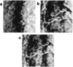
Macknik, Haglund, 1999. Article link.
|
|
|
Abstract: We optically imaged a visual masking illusion in primary visual cortex (area V-1) of rhesus monkeys to ask whether activity in the early visual system more closely reflects the physical stimulus or the generated percept. Visual illusions can be a powerful way to address this question because they have the benefit of dissociating the stimulus from perception. We used an illusion in which a flickering target (a bar oriented in visual space) is rendered invisible by two counter-phase flickering bars, called masks, which flank and abut the target. The target and masks, when shown separately, each generated correlated activity on the surface of the cortex. During the illusory condition, however, optical signals generated in the cortex by the target disappeared although the image of the masks persisted. The optical image thus was correlated with perception but not with the physical stimulus.
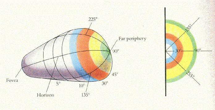
5. Activity in primary visual cortex predicts performance in a visual detection task.
Ress, D. Backus, B. T. & Heeger, D. J.

Subjects stared at a dot in the center of a screen while trying to anticipate correctly whether the image of a low-contrast ring would flash surrounding the dot. An fMRI mapped brain activity during this task. The following exerpt refers to V1 activity while the subjects were anticipating the ring image before the ring image appeared: “The base response was smaller in subregions of V1 corresponding to the central visual field and the far periphery than in subregions corresponding to the stimulus annulus. In other words, the pattern of V1 attention priming before sensory experience was shaped like the image they were anticipating. So something is painting a picture on V1, and the issue of visualization vs. attention may be moot for our purposes.
Visual Evoked Potentials:
6. Real-Time Analysis of Single Trial ERPs to Mediate Simple Communication in Sufferers of Locked-In Syndrome
C.R. Clark and N. Baum Psychology Discipline, Flinders University
Proceedings of the Inaugural Australasian Psychophysiology Conference, 1991
7. A Real-Time Brain-Computer Interface Based on Steady-State Visual Evoked Potentials
by
Rèmy Wahnoun et al. (Strangely, there is no date on this
document. Sometime after 2002?)
8. Identification of early VEP generators
(Clark et al., 1995, Human Brain Mapping) [circle of vixels, higher resolution]
9. Attention modulates responses in the human lateral geniculate nucleus
Daniel H. O'Connor et al. Nature Neuroscience 5,1203-1209 (2002)
10. Source Analysis of Event-related Cortical Activity during Visuo-spatial Attention
Francesco Di Russo, Antigona Martinez, Stephen A. Hillyard 2003
11. Cortical Sources of the Early Components of the Visual Evoked Potential
Francesco Di Russo, et al. 2001.
12. Identification of Early Visual Evoked Potential Generators by Retinotopic and Topographic Analyses
Vincent P. Clark, Silu Fan, and Steven A. Hillyard. Human Brain Mapping, 1995
V1 Mapping:
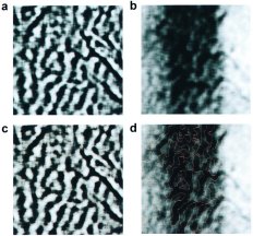
12. Functional analysis of primary visual cortex in humans
by Roger B.H. Tootell, et al. 1998. ( Excellent fMRI-based images of how visual field stimulation maps to V1.)
(Under construction) I am currently reading these and may add them later:
Selective attention gates visual processing in the extrastriate cortex.
Moran, J. and Desimone, R.
Attentional modulation of neural processing of shape, color and velocity in humans. Science 248
Corbetta, M., Miezin, F. M etc.
Visual, presaccadic, and congnitive activation of single neurons in monkey lateral intraparietal area
Colby, C. L., Duhamel, J. R. etc.
Neural Mechanisms of Spatial Selective Attention in Areas V1, V2,and V4 of Macaque Visual Cortex
Steven J. Luck, Leonardo Chelazzi, Steven A. Hillyard, and Robert Desimone ***